Otoplasty Techniques
There are several techniques for otoplasty (ear reshaping) that are used to help reshape the ear. Each technique is used for a different aspect in ear reshaping.
Mustarde Suture
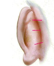
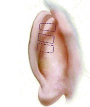
 The Mustarde suture is one of the most powerful techniques used in otoplasty. It is used when the antihelical fold is under developed, which causes the top portion of the ear to stick out further. When the top portion of the ear is not folded, the ear will look much larger and prominent than an ear with an appropriately developed antihelical fold. A good analogy is to think of a shirt collar. When it is unfolded, the collar sticks out further and can look expansive. However, with a fold within a collar, the relative size of the collar is diminished, making the collar look nearly half as prominent.
Mustarde sutures are performed by recreating the antihelix. Classically, this technique involves using a horizontal mattress suture which can help create the fold. Surgeons should be aware of the proper amount of cartilage to fold, as an overly prominent antihelical fold is a common postoperative complaint of an otoplasty. When recreated appropriately, the relative size of the ear as well as the distance of the ear from how far it sticks out from the head will be improved.
The preferred suture for Dr. Shah is the use of non-absorbable braided suture. Non-braided sutures can tear cartilage and unravel easily, which can cause the ear to look under corrected.
 Conchal Bowl Resection and Setback
The overly prominent conchal bowl is one of the most common complaints of patients with prominent ears. In this case, the central portion of the ear protrudes further than ideal. Here the surgeon must make the conchal bowl less prominent. In some cases, sutures to the conchal bowl to the mastoid periosteum (covering of the bone behind the ear) can help the middle portion of the ear look less prominent. In other cases, however, the conchal bowl is so large that simply suturing back is not going to improve the ear. A portion of the conchal bowl is resected and then sutured to the periosteum.
The preferred suture for here is also a non-absorbable braided suture.
 Caudal Helix and Lobule Prominence
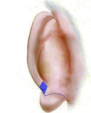
The overly prominent lower third of the ear can be result of a variety of factors including a prominently curved caudal helix. The caudal helix is the anatomic term for the lowest portion of the helical cartilage which extends to the ear lobule. In some cases, this cartilage can protrude causing the lobule to stick out. Correcting the lower prominence of the ear can be performed by either resecting the lower portion of the helical cartilage or suturing the lobule in a better position.


